

Tokyo researchers develop high-speed microscopy to video living cells
The new technique captures 3D images of biological processes at 19.2 frames per second, approximately 400 times faster than previous methods.
 Science Desk 2 min read
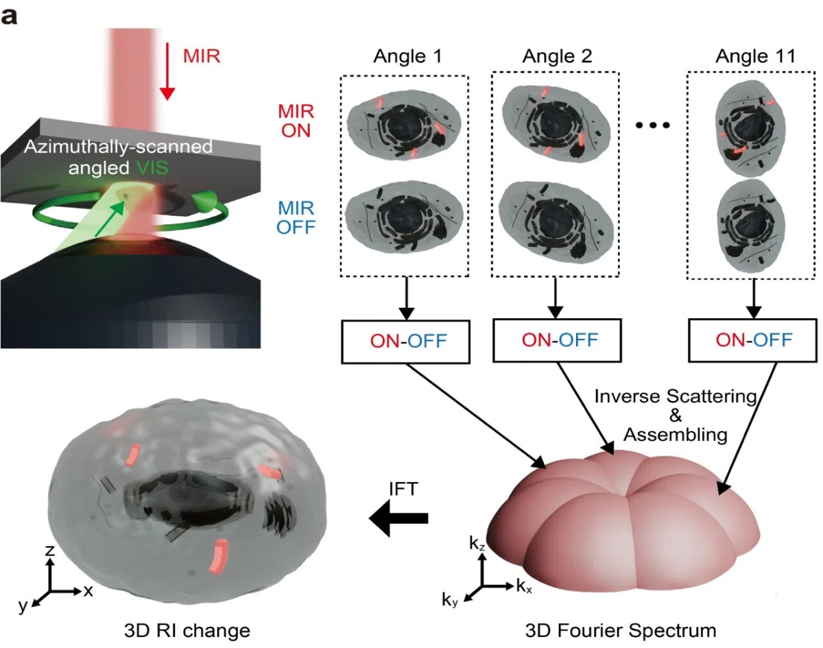
Researchers at the University of Tokyo have developed a high-speed microscopy technique that captures images quickly enough to make videos of dynamic processes inside living cells. The system, called video-rate mid-infrared photothermal optical diffraction tomography (MIP-ODT), can acquire a complete three-dimensional image every 52 milliseconds.
The team achieved a speed of 19.2 images per second, which is approximately 400 times faster than previous approaches that were limited to less than 0.05 images per second. To overcome the speed barrier, the researchers used a spatial light modulator to switch the illumination angle very quickly and maximized the signal-to-noise ratio for each shot.
The technique uses mid-infrared light to vibrate the chemical bonds within molecules, creating distinctive frequency signatures that can be used to map the internal composition of a cell. Unlike some other methods, this label-free approach uses no dyes, which can be toxic to live samples.
Using MIP-ODT, the team tracked how lipid droplets moved in three-dimensional space, revealing transport behaviors that could not be easily seen through 2D imaging alone. Masato Fukushima, the study's first author and a Ph.D. student in the Department of Physics, said the research transforms the technique from a method for observing relatively static samples into a tool for investigating fast biological processes in three dimensions.
The researchers foresee future applications for the technique, such as studying biomolecular condensates, which are membraneless structures containing proteins and nucleic acids. Because abnormal behavior of these condensates has been linked to neurodegenerative disorders, investigating how they change may help develop new treatments.
Senior author Takuro Ideguchi, a professor in the Graduate School of Frontier Sciences, said the ability to provide quantitative information about the chemical composition of living cells is a key strength. The team aims to use the technology to advance toward a quantitative, measurement-driven understanding of life.
Key facts
- The new microscopy technique captures 19.2 images per second.
- The technique is approximately 400 times faster than previous methods.
- The system uses mid-infrared light to create 3D images without using toxic dyes.
Three perspectives
Neutral
Researchers at the University of Tokyo have developed a high-speed microscopy technique to capture 3D images of living cells at 19.2 frames per second.
Positive
The new technique allows for the quantitative investigation of fast biological processes in three dimensions without using toxic dyes.
Negative
Capturing images at subsecond timescales has previously proven challenging due to the time required for signal averaging and sequential measurements.
Your week in news, every Wednesday
The biggest stories of the week for your edition, in your inbox every Wednesday morning, your time. Free, and you can unsubscribe at any time.